
Imagine your doctor says you need a closer look at your heart. They might order an ultrasound or an MRI. Both are powerful tools, but they work in completely different ways. One uses sound waves; the other uses strong magnets. Knowing the difference can help you understand what to expect and why one might be better for your specific situation.
We often hear these terms used interchangeably, but echocardiography is a non-invasive test that uses high-frequency sound waves to create moving images of the heart's chambers and valves. It’s like looking through a window into your chest while the heart beats in real-time. On the other hand, Cardiac MRI (or CMR) is an advanced imaging technique that uses magnetic fields and radio waves to produce highly detailed, three-dimensional pictures of heart structure and tissue health. While echo is quick and accessible, MRI offers a level of detail that is hard to beat.
How Do These Tests Actually Work?
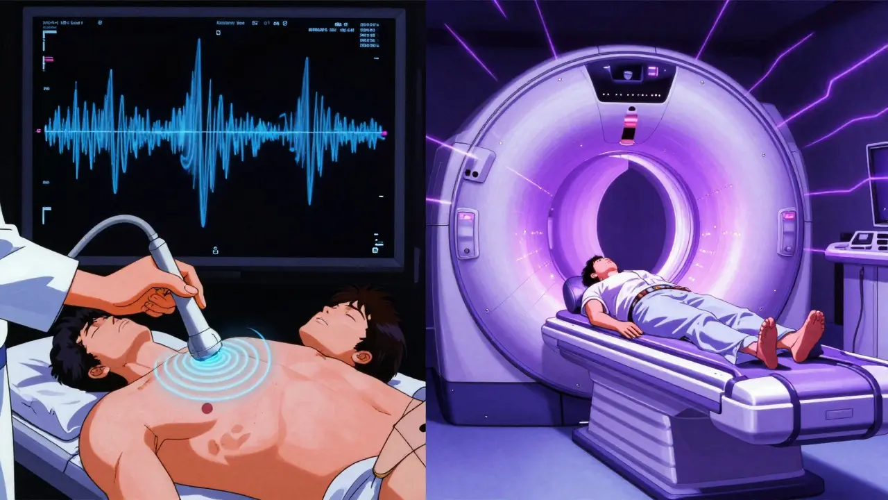
To get a clear picture, let's break down the mechanics. When you sit for an echocardiogram, a technician places a handheld device called a transducer on your chest. This device sends out sound waves-frequencies usually between 2 and 12 MHz-that bounce off your heart structures. The machine translates these echoes into video. Because it captures motion instantly, you can see your valves opening and closing right now. It’s fast, usually taking about 30 to 60 minutes, and involves no radiation.
Cardiac Magnetic Resonance Imaging works differently. You lie inside a large tube with a powerful magnet, typically 1.5 to 3.0 Tesla. Radio waves interact with the hydrogen atoms in your body’s tissues. The computer then builds a detailed image from this data. This process takes longer, often 45 to 90 minutes, because the machine has to capture multiple slices of the heart as it pauses briefly during each heartbeat. However, the result is a precise, three-dimensional map of your heart without relying on geometric assumptions.
Accuracy: Who Sees More Clearly?
If precision is your main concern, Cardiac MRI generally wins. Studies show that echo measurements can sometimes underestimate the size of your heart chambers or overestimate wall thickness. For example, research published in the Journal of Cardiovascular Magnetic Resonance found that echo often shows smaller ventricular dimensions compared to MRI. In cases where doctors need to measure exactly how much blood the heart pumps-the ejection fraction-MRI has lower variability between observers. A 2022 report noted that MRI’s inter-observer variability for ejection fraction is around 2.6%, whereas echo sits closer to 6.8%. This means two different doctors reading an MRI are more likely to agree on the numbers than two reading an echo.
However, echo isn’t “wrong.” It just relies on different methods. Standard 2D echo uses formulas to estimate volume based on shape, which can introduce errors if the heart isn't perfectly round. Newer 3D echo technology has narrowed this gap significantly. Still, when it comes to measuring heart mass or volume for conditions like heart failure, experts consider Cardiac MRI the gold standard.
| Feature | Echocardiography | Cardiac MRI |
|---|---|---|
| Imaging Method | Sound Waves (Ultrasound) | Magnetic Fields & Radio Waves |
| Average Cost (US) | $500 - $1,500 | $1,500 - $3,500 |
| Time Required | 30 - 60 Minutes | 45 - 90 Minutes |
| Tissue Detail | Good for structure/function | Excellent for tissue scarring/fibrosis |
| Real-Time Motion | Yes (High Frame Rate) | Limited by breath-holding |
| Accessibility | Widely available (Community Hospitals) | Less common (Specialized Centers) |
When Is Each Test Best Used?
Doctors don’t choose these tests randomly. They follow guidelines based on what they suspect is wrong. Echocardiography is the first-line tool for most initial checks. If you have shortness of breath, a heart murmur, or suspected valve disease, echo is usually the first stop. It’s portable, meaning it can even be brought to your bedside in the ICU. It’s also excellent for checking how well the right side of the heart is working in certain acute situations.
You’ll likely be referred for Cardiac MRI if the echo results are unclear or if the doctor needs to look deeper into the heart muscle itself. MRI is superior for detecting myocardial fibrosis (scarring), which is critical in diagnosing conditions like hypertrophic cardiomyopathy or cardiac sarcoidosis. It’s also the go-to for patients who are obese or have lung diseases that block ultrasound waves, creating what doctors call “poor acoustic windows.” In these cases, the sound waves can’t penetrate clearly, but the MRI sees right through.
What About Safety and Comfort?
Safety is a big part of the decision. Echo is incredibly safe. There are no known risks from the sound waves used. You can get an echo as many times as needed. It’s also quiet and open, so people with claustrophobia feel fine.
Cardiac MRI has more restrictions. The strong magnet means you cannot have certain metal implants, such as older pacemakers or some types of surgical clips. If you have an implant, your doctor must verify it is MRI-safe. Also, some MRIs use a contrast dye called gadolinium to highlight tissues. While generally safe, there is a small risk for people with severe kidney issues. The machine is also loud and enclosed, which can be stressful for those with anxiety or claustrophobia. However, newer low-field MRI machines (like 0.55T systems) are becoming available, which are quieter and more open, helping more patients qualify for the scan.

Cost and Availability: Can You Get the Test?
In the United States, about 15 million echos are performed annually, compared to 1.2 million cardiac MRIs. This huge difference comes down to access and cost. Echos are cheaper and available in almost every hospital and many clinics. You can often get one the same day if it’s urgent.
Cardiac MRI is more expensive, often costing twice as much as an echo. It also requires specialized equipment and trained technicians. According to recent reports, only about 35% of community hospitals offer same-week access to cardiac MRI, whereas nearly 80% have immediate echo availability. If your insurance covers it, the wait time for a non-urgent MRI can still exceed two weeks in some areas. This is why doctors start with echo-it’s faster, cheaper, and answers most basic questions.
The Future of Heart Imaging
Technology is blurring the lines between these two worlds. AI is being integrated into echo machines to reduce human error and make measurements more consistent with MRI standards. Meanwhile, MRI technology is getting faster and more accessible. Experts predict that by 2030, we will see more “hybrid” protocols where doctors use both tools together for complex cases. For now, though, echo remains the frontline scout, and MRI is the deep-dive specialist.
Which heart scan is better for detecting heart attacks?
For detecting old heart attacks and the amount of scar tissue left behind, Cardiac MRI is superior. It can precisely identify areas of dead or damaged heart muscle using late gadolinium enhancement. Echocardiography can show wall motion abnormalities but cannot visualize the scar tissue itself as clearly.
Can I have a Cardiac MRI if I have a pacemaker?
It depends on the type of pacemaker. Older devices are strictly contraindicated due to the strong magnetic field. However, many modern pacemakers and ICDs are labeled "MRI-conditional." Your cardiologist and electrophysiology team must verify your specific device model and program it safely before the scan.
Why did my doctor order an MRI after my echo was normal?
Sometimes an echo looks normal, but symptoms persist. An MRI can detect subtle changes in heart tissue, such as early inflammation (myocarditis) or infiltration (amyloidosis), that do not yet affect the heart's pumping function. It provides a higher level of diagnostic certainty when the initial screen is inconclusive.
Does an echocardiogram hurt?
No, a standard transthoracic echocardiogram is painless. It involves pressing a probe against your chest with gel. You may feel slight pressure, but no pain. It is non-invasive and does not involve needles or radiation.
How long do I have to hold my breath during a Cardiac MRI?
You will be asked to hold your breath for short periods, usually 10 to 20 seconds, repeatedly throughout the scan. This prevents motion blur from breathing. The technologist will guide you through these pauses. If you cannot hold your breath, newer techniques allow for free-breathing scans, though they take longer.